

When you’ve had a routine screening mammogram, the last thing you want to hear is that something suspicious was found. Fortunately, most abnormal mammograms do not end up as cancer.
During a screening mammogram, the technician and physician are looking for unusual breast changes, such as lumps, masses or small white spots, called calcifications. If this is not your first mammogram, your results will be compared to previous mammograms. That way, the doctor can note if any irregularities in your breast tissue were already there or if they are new and should be investigated.
If there is something suspicious, you will be called back for additional imaging and possibly other testing. It’s most likely nothing, but if it is cancer, the goal is to find it at the earliest, most treatable stage.
What else could it be?
There are several reasons that you may be called back after a mammogram and it doesn’t necessarily mean you have cancer. For example, some women naturally have denser breast tissue, making it harder to get a good image during routine mammogram. You may also have a cyst or other mass that isn’t cancer. Sometimes, the doctor just wants a closer look, because the first image was unclear.
What happens next?
If you are called back after a mammogram, or if you or your doctor found a lump in your breast during manual exam, your doctor will order a diagnostic mammogram. This is the same type of imaging as a screening mammogram, except more pictures may be taken. A radiologist will be reading the images in real time, so any additional imaging can be done on the same visit.
Other possible tests you may need include:
- 3-D mammogram — Similar to a CT scan, to take multiple views and create a 3-D picture of your breast. This type of mammogram allows doctors to see finer details more clearly and is better at detecting breast cancer in women with dense breast tissue.
- Breast MRI — A breast MRI can detect cancers that mammograms and ultrasound may miss, or more closely identify non-cancerous cysts and masses.
- Breast ultrasound — Breast ultrasound uses high-frequency sound waves to acquire images of your breast tissue on a computer screen. This test may be used in conjunction with mammogram.
- Breast biopsy — A needle biopsy may be recommended for lesions found on imaging. During biopsy, your doctor will remove a small sample of your breast tissue under image guidance (ultrasound, MRI, or mammogram). The sample is then examined under a microscope to see if it is cancerous. A biopsy is the only way to know if cells are cancer.
Once you’ve had any of the additional diagnostic tests ordered, your doctor will call you with the results. In many cases, the extra tests will confirm that your breast tissue is normal and that will be the end of your worry.
What if it’s cancer?
If it turns out that your results show you have breast cancer, a doctor will refer you to a breast surgeon. She will discuss your next steps with you and answer any questions you may have. The earlier breast cancer is detected, the better chance that treatment will be successful.
Learn more about breast health at The Christ Hospital and make an appointment with one of our women’s health experts! Schedule your screening mammogram online or by calling 513-585-2668.
Learn more about breast health at The Christ Hospital and make an appointment with one of our women’s health experts! Schedule your screening mammogram online or by calling 513-585-2668.
Schedule a Mammogram
Featured in This Post
 Hamilton County health officials report more cyclosporiasis cases - opens in a new tab - external linkHamilton County health officials have reported more new cyclosporiasis cases. Health officials also say the county is seeing a "statistically significant increase" in people seeking care for gastrointestinal issues, regardless of the cause.
 Christ Hospital book fair - opens in a new tab - external linkThe Christ Hospital Health Network and Joseph-Beth Booksellers hosted a Buy a Book, Give a Book fair in support of local students through United Way of Greater Cincinnati’s Backpacks for Success initiative.
 Electrolyte packets claim to hydrate faster than water. Do you actually need them? - opens in a new tab - external linkElectrolyte packets have become a popular choice for staying hydrated, especially during the summer months. But do you actually need them? We took that question to the experts.
 The Christ Hospital Health Network to Host Free Webinar on Heart ConditionsFree Lunch and Learn Webinar on Thursday, July 30 from Noon – 1:00 p.m.
- Conversations for Confidence: Expert Answers About Spine CareIf you have back or neck pain, confusion and uncertainty about treatment options can add to your frustration. Here are some answers from our experts to help you find relief.
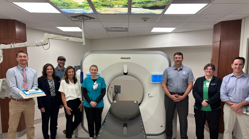 Christ Hospital Health Network first in country to install, use new CT scanner for radiation treatment - opens in a new tab - external linkThe Christ Hospital Health Network has installed and begun use of Philips' new Rembra Radiation Therapy (RT) Computed Tomography (CT) system to support efficient cancer treatment planning workflows.
- The Christ Hospital Health Network First in North America to Install Philips Newest CT Solution for Radiation TherapyPhilips Rembra RT Advanced CT Scanner Now in Clinical Use, Supporting More Precise and Efficient Radiation Therapy Planning Workflows
 The Christ Hospital Health Network, Joseph-Beth Partner with United Way to Host Book Fair to Help Local Students Start School Ready to LearnA Buy a Book, Give a Book campaign will support the Backpacks for Success initiative
